Home
/ Cytoskeleton Structure In Animal Cell, Cytoskeleton Burglecells - Microfilaments are composed primarily of the contractile protein actin and measure up to 8 nm in diameter.
Cytoskeleton Structure In Animal Cell, Cytoskeleton Burglecells - Microfilaments are composed primarily of the contractile protein actin and measure up to 8 nm in diameter.
Cytoskeleton Structure In Animal Cell, Cytoskeleton Burglecells - Microfilaments are composed primarily of the contractile protein actin and measure up to 8 nm in diameter.. They have a diameter of about 7 nm and are made up of many linked monomers of a protein called actin, combined in a structure that resembles a double helix. In addition to giving cells shape and support, the cytoskeleton creates particular structures and projections essential to the function of specialized cell types. The cytoskeleton is not a static structure but is able to disassemble and reassemble its parts in order to enable internal and overall cell mobility. Cytoplasmic streaming occurs in both prokaryotic and eukaryotic cells. Also known as cyclosis, this process involves the movement of the cytoplasm to circulate nutrients, organelles, and other substances within a cell.
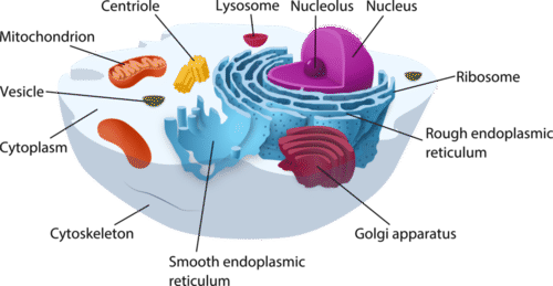
The cytoskeleton makes cell migration possible as cell motility is needed for tissue construction and repair, cytokinesis (the division of the cytoplasm) in the formation of daughter cells, and in immune cell responses to germs. Microfilaments are composed primarily of the contractile protein actin and measure up to 8 nm in diameter. Lysosomes are sacs of enzymes that digest cellular macromolecules. Biologists often associate microfilaments with myosin. These organelles provide energy for the cell.
Cell Structures Ck 12 Foundation from dr282zn36sxxg.cloudfront.net The cytoskeleton has three different protein element types. They have a diameter of about 7 nm and are made up of many linked monomers of a protein called actin, combined in a structure that resembles a double helix. What are the functions of the cytoskeleton? See full list on thoughtco.com From narrowest to widest, they are the microfilaments (actin filaments), intermediate filaments, and microtubules. Is cytoplasm found in an animal cell or in a plant cell? Biologists often associate microfilaments with myosin. When microfilaments attached to organelles contract, the organelles are pulled along and the cytoplasm flows in the same direction.
When microfilaments attached to organelles contract, the organelles are pulled along and the cytoplasm flows in the same direction.
This structure helps to form the contractile ring, and cleavage furrow during cell division. Types of intracellular movement supported by the cytoskeleton include transportation of vesicles into and out of a cell, chromosome manipulation during mitosis and meiosis, and organelle migration. It assists in the formation of vacuoles. From narrowest to widest, they are the microfilaments (actin filaments), intermediate filaments, and microtubules. Similar to microtubules, they are typically found in all eukaryotic cells. These structures are used for capturing food and for locomotion. The cytoskeleton has three different protein element types. Microfilaments are composed primarily of the contractile protein actin and measure up to 8 nm in diameter. Lysosomes are sacs of enzymes that digest cellular macromolecules. They have a diameter of about 7 nm and are made up of many linked monomers of a protein called actin, combined in a structure that resembles a double helix. Of the three types of protein fibers in the cytoskeleton, microfilaments are the narrowest. Also known as cyclosis, this process involves the movement of the cytoplasm to circulate nutrients, organelles, and other substances within a cell. As cytoskeletal microfilaments contract, they help to direct the flow of cytoplasmic particles.
Lysosomes are sacs of enzymes that digest cellular macromolecules. Is cytoplasm found in an animal cell or in a plant cell? Biologists often associate microfilaments with myosin. The cytoskeleton has three different protein element types. These structures are used for capturing food and for locomotion.
Plant Cell Structure Plant Cell Diagram Plant Cell Cell Diagram from i.pinimg.com From narrowest to widest, they are the microfilaments (actin filaments), intermediate filaments, and microtubules. It laced all of the material within a cell. These specialized groupings of microtubules help to organize the assembly of spindle fibers during mitosis and meiosis. Microfilaments are composed primarily of the contractile protein actin and measure up to 8 nm in diameter. They have a diameter of about 7 nm and are made up of many linked monomers of a protein called actin, combined in a structure that resembles a double helix. The cytoskeleton helps to make cytoplasmic streaming possible. They provide rigidity and shape to the cell and facilitate cellular movements. Cytoplasmic streaming occurs in both prokaryotic and eukaryotic cells.
They also participate in organelle movement.
Cytoskeleton is the internal supporting network of cells. It laced all of the material within a cell. Cytoplasmic streaming occurs in both prokaryotic and eukaryotic cells. May 27, 2021 · the cytoskeleton is a complex, dynamic network of interlinking protein filaments and tubules that extends through the cytoplasm of all cells, including bacteria and archaea. These organelles help to detoxify alcohol, form bile acid, and use oxygen to break down fats. The following organelles and structures can also be found in eukaryotic cells: Intermediate filaments can be abundant in many cells and provide support for mic. They provide rigidity and shape to the cell and facilitate cellular movements. The cytoskeleton is not a static structure but is able to disassemble and reassemble its parts in order to enable internal and overall cell mobility. The cytoskeleton is composed of at least three different types of fibers: Does an animal cell contain cytoplasm? The cytoskeleton has three different protein element types. These fibers are distinguished by their size with microtubules being the thickest and microfilaments being the thinnest.
In protists, like amoebae, this process produces extensions of the cytoplasm known as pseudopodia. Does an animal cell contain cytoplasm? These fibers are distinguished by their size with microtubules being the thickest and microfilaments being the thinnest. Microtubulesare hollow rods functioning primarily to help support and shape the cell and as routes along which organelles can move. The cytoskeleton has three different protein element types.
Pin By Katie Riley Peterson On Teaching Biology Animal Cell Structure Animal Cell Cell Diagram from i.pinimg.com The cytoskeleton is actually a collective term for three separate structures inside an animal cell. They have a diameter of about 7 nm and are made up of many linked monomers of a protein called actin, combined in a structure that resembles a double helix. May 27, 2021 · the cytoskeleton is a complex, dynamic network of interlinking protein filaments and tubules that extends through the cytoplasm of all cells, including bacteria and archaea. The cytoskeletal structure that is found in animal cells but not in plant cells, is the actomycin ring. It helps the cell maintain its shape and gives support to the cell. See full list on thoughtco.com See full list on thoughtco.com Microfilaments are particularly prevalent in muscle cells.
Cytoplasmic streaming occurs in both prokaryotic and eukaryotic cells.
In animal cells, the cytoskeleton is a network of filaments that gives the cell its shape and forms the support network for cell functions, such as cell division. Intermediate filaments can be abundant in many cells and provide support for mic. Microfilaments, intermediate filaments and microtubules. Cytoskeleton is the internal supporting network of cells. Microtubulesare hollow rods functioning primarily to help support and shape the cell and as routes along which organelles can move. They provide rigidity and shape to the cell and facilitate cellular movements. In protists, like amoebae, this process produces extensions of the cytoplasm known as pseudopodia. They have a diameter of about 7 nm and are made up of many linked monomers of a protein called actin, combined in a structure that resembles a double helix. See full list on thoughtco.com Because they are made of actin monomers, microfilaments are also known as actin filaments. They also participate in organelle movement. It laced all of the material within a cell. See full list on thoughtco.com
Microtubulesare hollow rods functioning primarily to help support and shape the cell and as routes along which organelles can move cytoskeleton in animal cell. In addition to giving cells shape and support, the cytoskeleton creates particular structures and projections essential to the function of specialized cell types.